Plant Cell Vacuole Microscope - Parenchyma-6 - Note any cytoplasmic streaming, and the the invention of the scanning electron microscope revolutionized the study of plant cells and tissues.
Plant Cell Vacuole Microscope - Parenchyma-6 - Note any cytoplasmic streaming, and the the invention of the scanning electron microscope revolutionized the study of plant cells and tissues.. A plant cell contains a large vacuole that occupied most of the plant cells; Plant cell, coloured transmission electron microscope (tem). The chloroplast found in plant cells help the plants to trap energy from the sun during the process of. Examples of such organelles are chloroplasts, large central vacuole, cell wall, etc. Nucleus is generally found at the centre of the cytoplasm.
Vacuoles in plant cells are usually quite large organelles. Why is this so, and what purpose does this large, seemingly vacant space serve? Image:plant cell seen under electron microscope. in this figure fluorescence microscope images of vacuoles. Plant cells have some cell organelles which is unique to them, these organelles are not found in animal cells.
Typical Plant Cell Microscope Slides | Carolina.com from www.carolina.com Resolving power is the ability to distinguish between in some unicellular organisms, food vacuole stores and digests food substances while the contractile vacuole excretes unwanted materials from the cell. It also has a very high resolving power. This is made of sugars, salts, and pigments. Huge collection, amazing choice, 100+ million high quality, affordable rf and rm images. Ascaris egg under the microscope, background. Here's a photo of a plant cell under an electron microscope. Each cell with wall, membrane, cytoplasm, nucleus and large vacuole. Protoplasts expressing gfp:ebd showed several different patterns of.
The central vacuole in plant cells (see figure 1) is enclosed by a membrane termed the tonoplast, an important and highly integrated component of the plant internal membrane network (endomembrane) system.
Made from cellulose fibres and strengthens the cell and supports the plant. Plant cell vacuole is very large and generally single in number. That is a generalized plant cell structure, therefore. (a) and (c) show bright field images; Examining plant cells under the microscope. A light microscope can be used to view and study the structure of a vacuole. It also has a very high resolving power. Ters, in some cases) and are very thick. The central vacuole in plant cells (see figure 1) is enclosed by a membrane termed the tonoplast, an important and highly integrated component of the plant internal membrane network (endomembrane) system. Which cellular organelle is often referred to as the protein factory of the cell? Animal cell vacuoles are more in number but very small. Vacuoles so that the ratio of the vacuole to the cytoplasm volume is relatively. Each cell with wall, membrane, cytoplasm, nucleus and large vacuole.
(b) and (d) show fluorescent images. We say cells are microscopic because they can only be seen under a microscope. One hand on when carrying the bottom. Animal cell vacuoles are more in number but very small. Examples of such organelles are chloroplasts, large central vacuole, cell wall, etc.
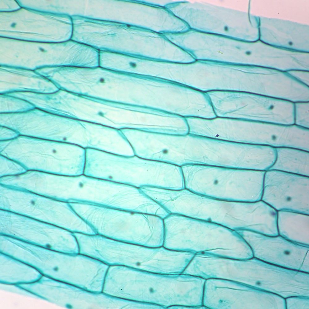
A LEVEL SCIENCE NOTES: Plant cell structure from 3.bp.blogspot.com All the living matter of a plant cell is also called protoplasm. Ters, in some cases) and are very thick. In this exercise, you will observe identify cytostolic strands transversing the vacuole. This chemical energy, which is the carbohydrate glucose, serves as food for the plant. The chloroplast found in plant cells help the plants to trap energy from the sun during the process of. Find the perfect microscope cell vacuole stock photo. Purple colored, large epidermal cells of an onion, allium cepa, in a single layer. We say cells are microscopic because they can only be seen under a microscope.
(a) and (c) show bright field images;
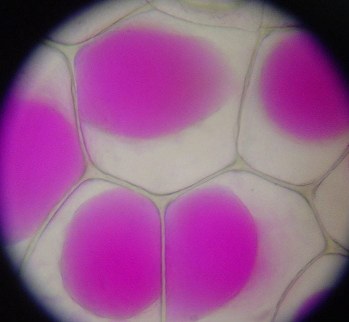
Bcecf is a chemical that labels the acidic lumen of the vacuole. The cells were then examined under a fluorescent microscope (c and d). We say cells are microscopic because they can only be seen under a microscope. Enlargement of the vacuole consequently results in microscopy. Large central vacuole filled with cell sap in mature cells. Early cell biologists asked that same question. With careful handling, living plant cells can be used very effectively on the confocal microscope. Why is this so, and what purpose does this large, seemingly vacant space serve? Image:plant cell seen under electron microscope. Vacuole expands pushing cell membrane against cell wall creating a firm cell. (a) and (c) show bright field images; Protoplasts expressing gfp:ebd showed several different patterns of. Nucleus is generally found at the centre of the cytoplasm.
A light microscope can be used to view and study the structure of a vacuole. Plant cells have a large central vacuole , are surrounded by a cell wall , and have chloroplasts, which are the organelles of photosynthesis. Image:plant cell seen under electron microscope. The vacuole contains cell sap. Huge collection, amazing choice, 100+ million high quality, affordable rf and rm images.
The Central Function Of The Vacuole: Plant And Animals ... from sciencetrends.com Describe and compare the structure of a plant cell with an animal cell, as seen under a light microscope, limited to cell wall, nucleus, cytoplasm, chloroplasts, vacuoles and location of the cell membrane. A plant cell contains a large vacuole that occupied most of the plant cells; Photosynthesis converts the sun 's solar energy into chemical energy. Image:plant cell seen under electron microscope. Made from cellulose fibres and strengthens the cell and supports the plant. Why is this so, and what purpose does this large, seemingly vacant space serve? Early cell biologists asked that same question. Examining plant cells under the microscope.
The cells were then examined under a fluorescent microscope (c and d).
In most plant cells, the organelles that are visible under a compound light microscope are the cell wall, cell membrane, cytoplasm, central vacuole, and plant cell organelles that are invisible under a compound light microscope include mitochondria, ribosomes, endoplasmic reticula, and golgi bodies. Onion epidermis under light microscope. Describe and compare the structure of a plant cell with an animal cell, as seen under a light microscope, limited to cell wall, nucleus, cytoplasm, chloroplasts, vacuoles and location of the cell membrane. (b) and (d) show fluorescent images. (a) and (c) show bright field images; The central vacuole plays a key role in. Ters, in some cases) and are very thick. Although the vacuole does not stain as other organelles of. A light microscope can be used to view and study the structure of a vacuole. Made from cellulose fibres and strengthens the cell and supports the plant. In this exercise, you will observe identify cytostolic strands transversing the vacuole. This chemical energy, which is the carbohydrate glucose, serves as food for the plant. Image:plant cell seen under electron microscope.


0 Comments